
Plant Stress Biology Surveillance and Experimental Design
Sara Laranjeira
PheNo coordinator
Revealing the world of color to future plant phenomicists
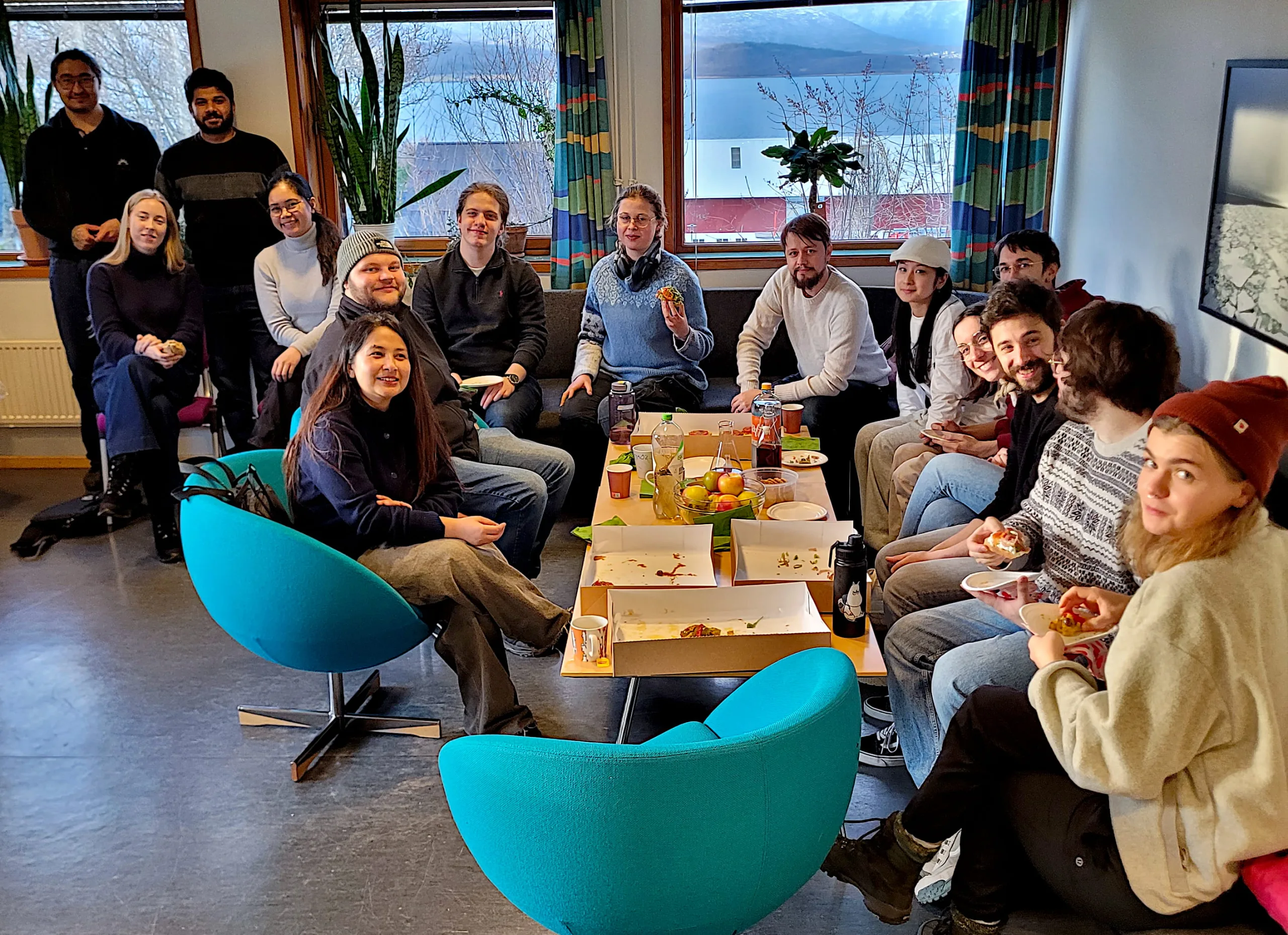
At this time of the year, the night sky over Tromsø is usually draped in shades of green and red from the northern lights. However, because of cloudy weather, our students had to find colors elsewhere; more specifically, in our course BIO8033 – Plant Stress Biology Surveillance and Experimental Design. An intensive but successful course with eight PhD and six MSc students, from NTNU (Trondheim/Gjøvik), NMBU (Ås), the University of Helsinki, and UiT.
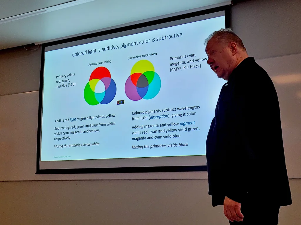
This November, last week, UiT – Arctic University of Norway (Tromsø) organized the third edition of BIO8033/3033 for PhD and MSc students, on behalf of Photosyntech, PheNo, and NordPheno. Professor Markku Keinänen (University of Eastern Finland) led the course, which included lectures on color and imaging theory, how to use it in practical phenotyping applications, and hands-on data-processing sessions.
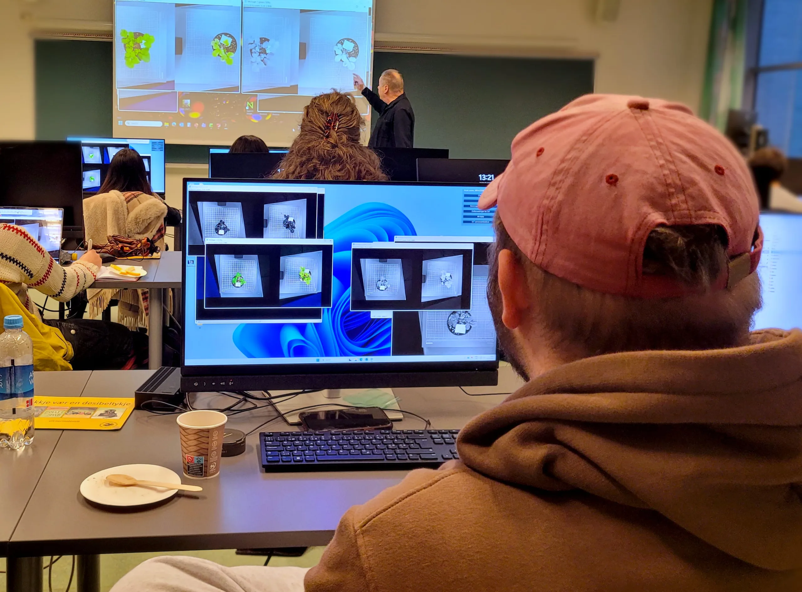
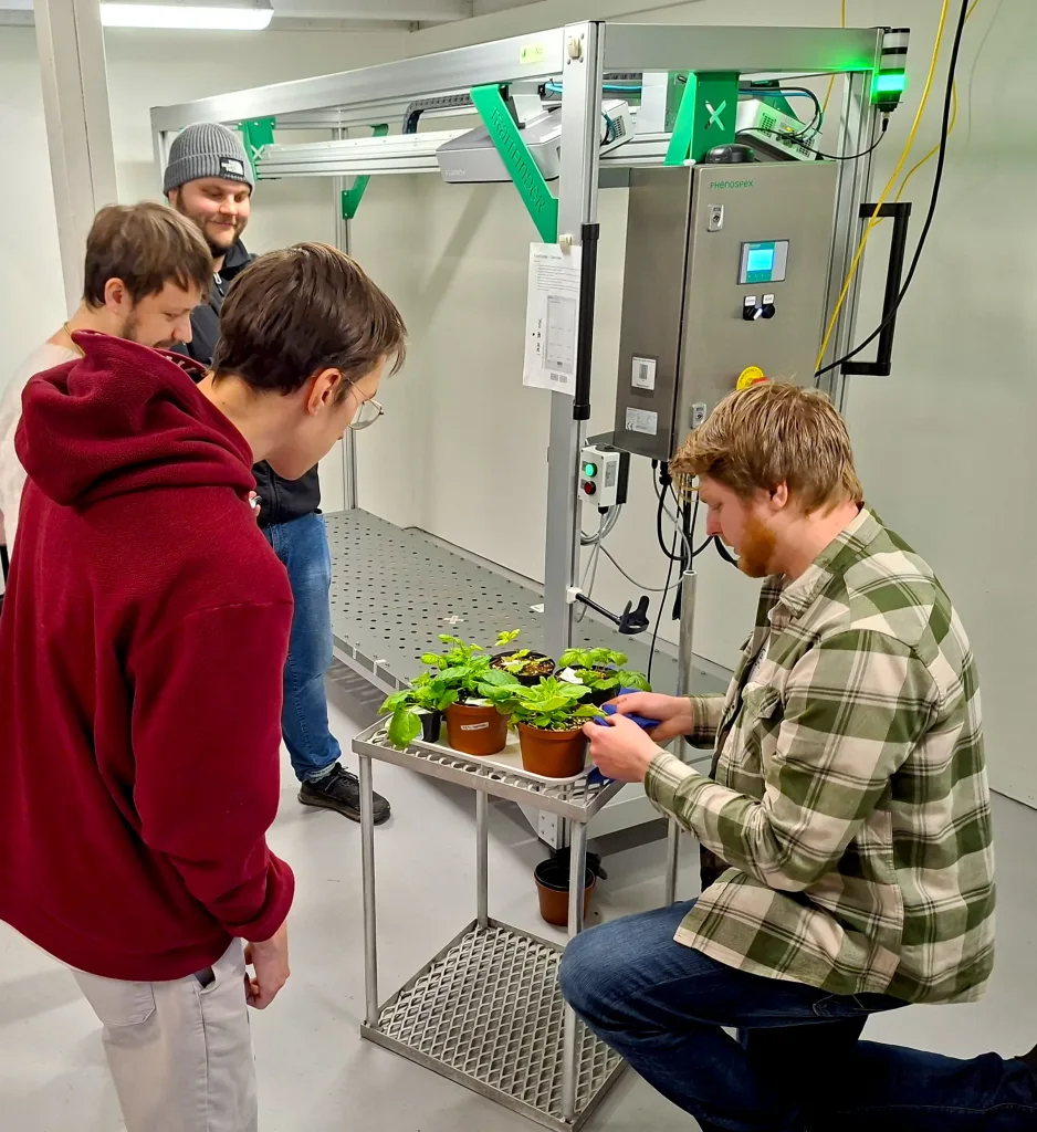
The students collected their own imaging data at the Climate Laboratory – Holt, where a short stress-treatment experiment had been conducted. The data acquisition gave the students an opportunity to see and use the available imaging equipment, including hyperspectral cameras, a 3D multispectral scanner (TraitFinder, PlantEye), a thermal camera, and an RGB-phenotyping station. The images were taken from commonly used model plants under experimental treatments of drought or high-light intensity stress.
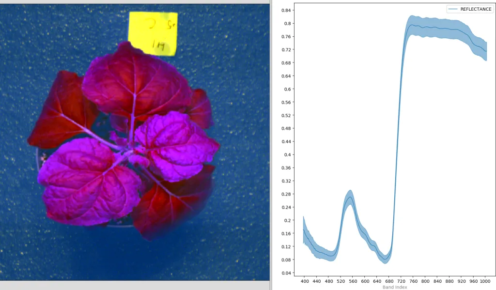
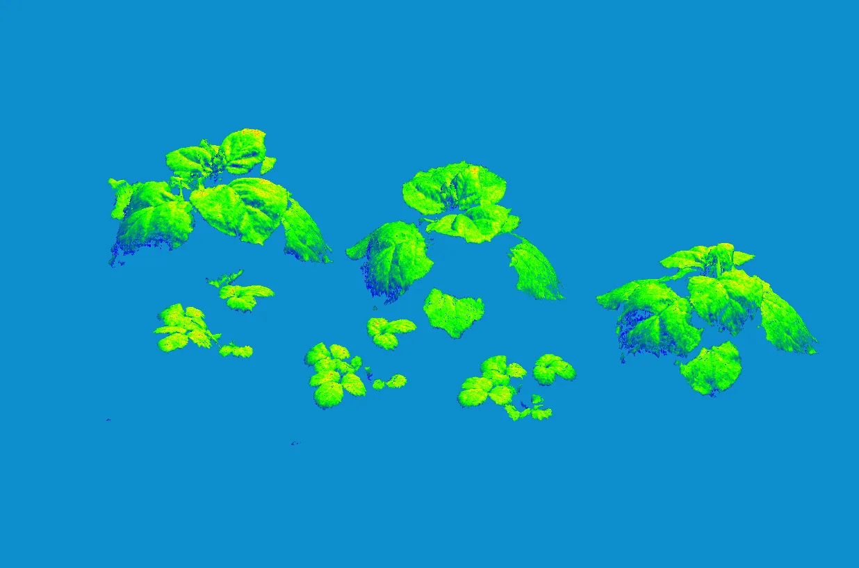
The hands-on data-processing sessions focused on data exploration and showed students that an image can include much more than what the naked eye can detect. They learned how to use specific software commonly used in phenotyping workflows, including Spectronon, CloudCompare, and Fiji/ImageJ. Here, they learned that there are many considerations one needs to take both when capturing and processing imaging data originating from multiple sensors and with considerable variation in file sizes and computing requirements. The students were shown that color can be numerically represented in a plethora of ways, to enhance features like signs of stress in plant tissue. Given the sensors available for the course, it was also possible to explore spectral signatures outside the visible spectrum, into the infrared region, and digital 3D models used to non-invasively calculate morphological parameters.
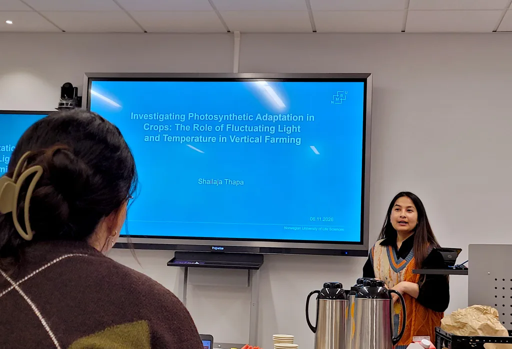
The course wrapped up with a seminar, where each student presented a paper on a phenotyping project, using relevant and similar equipment used earlier during the week. Some students opted to present plans for their PhD project, and received some feedback linking their local phenotyping equipment with the course content. The latter should fit nicely into the students’ final report on setting up and conducting a hypothetical phenotyping project utilizing imaging techniques.
The course was yet another example of how important it is to get together to share ideas and insight on how to run a project, as information flowed freely between students and lecturers. With students originating from universities across Norway, as well as other Nordic countries, tighter bonds between our research groups were formed.
We would like to thank all participants, organizers, and instructors for making the course a success. Special thanks to Markku Keinänen and Marius Dobbe Klemetsen for the lectures and data lab sessions; Laura Jaakola and Katja Karppinen for organizing the course; Wojciech Leoniuk for coordinating the experimental activities; and Sara Pastacaldi and Vasili Balios for leading the RGB and thermal imaging stations, respectively.